
*以下内容中所有出现的LCI指联动成像技术 (LCI: Linked Color lmaging); BLI指蓝光成像技术 (BLI: Blue Light lmaging)
王晔主任从以下几个方面进行讲解:
- 内镜成像技术的发展史
- BLI和LCI的技术原理
- LCI对食管,胃和结肠的临床应用优势
以下内容为部分摘要,完整内容敬请点击视频观看!
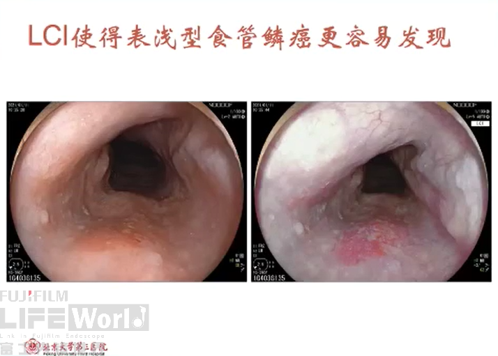
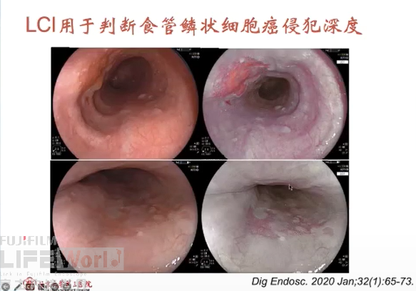
LCI在食管的临床应用:IIb型的早期食管癌往往很难被发现,而LCI突出了色彩的变化,更容易诊断
有研究文献证明:
1. LCI更容易发现表浅型食管鳞癌1;
2.能够区分食管鳞癌与上皮内瘤变2;
3.判断食管鳞状细胞癌侵犯深度3;

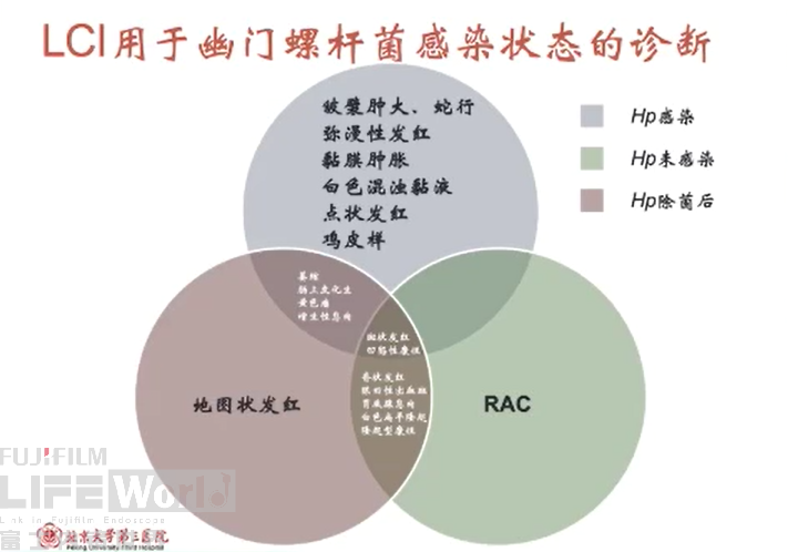
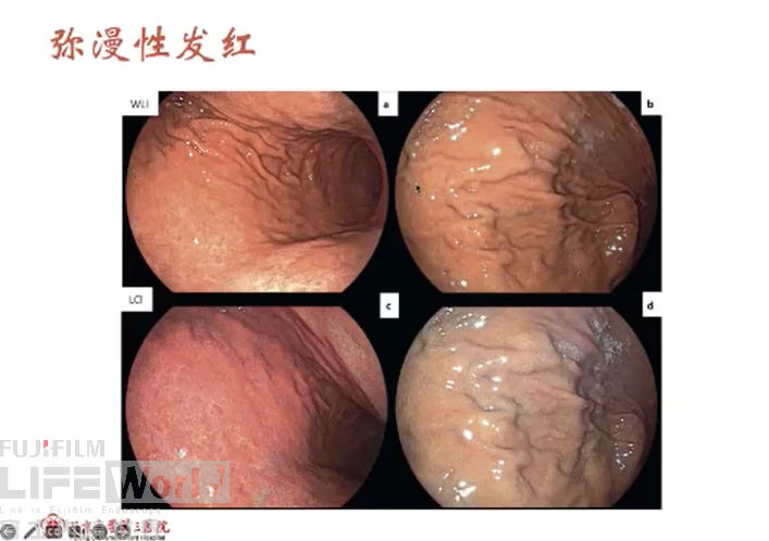
LCI在胃的临床应用:胃镜除了发现早癌,还可以判断患者胃癌的风险,特别是幽门螺杆菌感染状态。根据京都胃炎分类,不同内镜下的表现,我们可以判断患者处于哪个状态(HP未感染/HP感染/HP除菌后)
有研究文献证明:
1. LCI下对于幽门螺旋杆菌感染诊断率高于白光,特别是除菌后状态的诊断4
2. LCI下对于早期胃癌病灶范围判断的准确率明显优于白光5
3. 在日常胃镜筛查下,超细内镜结合LCI模式判断早癌的阳性率优于白光内镜6
4. LCI模式下,诊断除菌后的胃癌漏诊率明显低于白光内镜7
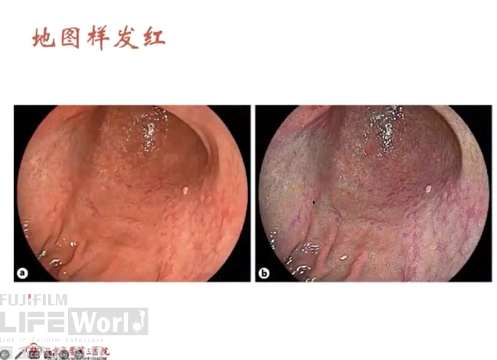
LCI在结肠的临床应用:可预测结肠息肉的病理类型
有研究文献证明:
1. 和白光内镜相比,LCI预测病理类型敏感性和特异性更高,不同观察者的一致性更高。增生性息肉颜色更偏白(接近于背景黏膜),腺瘤黏膜更红,LCI下色彩差异更明显8
2. LCI下区分腺瘤、无蒂锯齿状息肉、增生性息肉和非增生性息肉准确度更高9
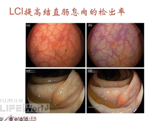
3. LCI能够提高结直肠息肉的检出率,特别对于<10mm的微小息肉差异性更明显10
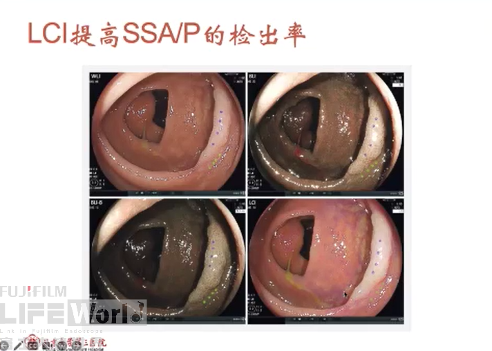
4. LCI能够提高SSA/P检出率,特别是平坦型病变,无蒂锯齿状病变11
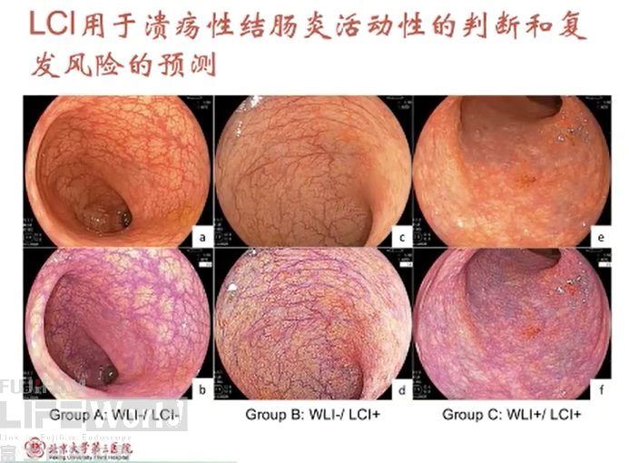
5. LCI可判断溃疡性结肠炎的活动性和预测复发风险12
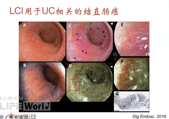
6. LCI 判断UC相关的结直肠癌的检出率更高13

文献出处
[1] Nakamura, Koki, et al. "Usefulness of linked color imaging in the early detection of superficial esophageal squamous cell carcinomas." Esophagus 18 (2021): 118-124.
[2] Tsunoda, Masato, et al. "New diagnostic approach for esophageal squamous cell neoplasms using linked color imaging and blue laser imaging combined with iodine staining." Clinical Endoscopy 52.5 (2019): 497-501.
[3] Kobayashi, Kenichi, et al. "Color information from linked color imaging is associated with invasion depth and vascular diameter in superficial esophageal squamous cell carcinoma." Digestive Endoscopy 32.1 (2020): 65-73.
[4] Dohi, Osamu, et al. "Linked color imaging improves endoscopic diagnosis of active Helicobacter pylori infection." Endoscopy international open 4.07 (2016): E800-E805.
[5] Fockens, Kiki, et al. "Linked color imaging improves identification of early gastric cancer lesions by expert and non-expert endoscopists." Surgical Endoscopy 36.11 (2022): 8316-8325.
[6] Khurelbaatar, Tsevelnorov, et al. "Improved detection of early gastric cancer with linked color imaging using an ultrathin endoscope: a video-based analysis." Endoscopy International Open 10.05 (2022): E644-E652.
[7] Kitagawa, Yoshiyasu, et al. "Comparison of endoscopic visibility and miss rate for early gastric cancers after Helicobacter pylori eradication with white‐light imaging versus linked color imaging." Digestive Endoscopy 32.5 (2020): 769-777.
[8] Min, Min, et al. "Development and validation of the linked color imaging classification for endoscopic prediction of colorectal polyp histology." Journal of Digestive Diseases 23.5-6 (2022): 310-317.
[9] Houwen, Britt BSL, et al. "Real-time diagnostic accuracy of blue light imaging, linked color imaging and white-light endoscopy for colorectal polyp characterization." Endoscopy International Open 10.01 (2022): E9-E18.
[10] Wang, Jun, et al. "The Effect of Linked Color Imaging for Adenoma Detection. A Meta-analysis of Randomized Controlled Studies." Journal of Gastrointestinal & Liver Diseases 31.1 (2022).
[11] Li, Jun, et al. "Colorectal sessile serrated lesion detection using linked color imaging: A multicenter, parallel randomized controlled trial." Clinical Gastroenterology and Hepatology 21.2 (2023): 328-336.
[12] Matsumoto, Kenta, et al. "Clinical usefulness of linked color imaging for evaluation of endoscopic activity and prediction of relapse in ulcerative colitis." International Journal of Colorectal Disease 36 (2021): 1053-1061. [13] Kanmura, Shuji, et al. "A case of screening colonoscopy using linked-color imaging to detect ulcerative colitis–associated colorectal cancer." Digestive and Liver Disease 51.7 (2019): 1061.
讲课出处:中国医药卫生事业发展基金会消化专家委员会第二届年会